
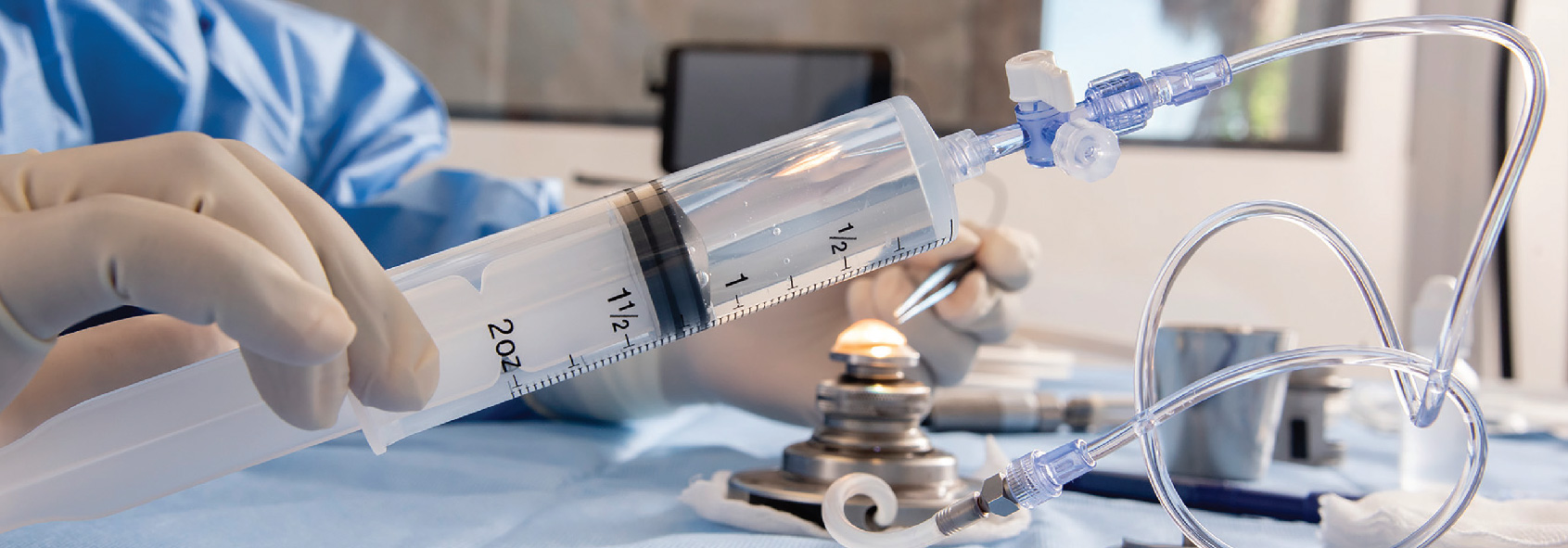
Advanced Cornea Processing
Advanced Cornea Processing for DMEK
Hydrodissection “Blister” Technique
- No-Touch Technique
- Isolates exposure of Trypan Blue to DM
- Instant staining during dissection
- Graft is supported evenly by fluid during dissection
- Decreases endothelial cell loss
- Decreases failure rates
LWVI introduced the Blister technique in 2012 after multiple internal studies demonstrated this unique procedure safer and more reliable for gently dissecting DMEK grafts. Most eye banks continue to use the “Submerged Cornea Using Backgrounds Away” (SCUBA) method because it is the initial approach used by surgeons in the operating room. The advantages of the Blister method align with LWVI’s innovative culture of improving standards and providing the highest quality tissue possible with the objective of improving clinical outcomes.
A Game Changer

“Having peeled close to a thousand DMEK donors with the SCUBA technique has given me a unique perspective on the tremendous advantages of the hydrodissection (blister) technique.
I only use the Blister Method. It’s a game changer!”
— Mark Gorovoy, MD
Gorovoy MD Eye Specialists, Fort Myers, Florida
Advanced Cornea Processing for UT-DSAEK
High-Pressure Anterior Chamber (HPAC) Technique
High-Pressure Anterior Chamber (HPAC), LWVI’s proprietary technique for preparing ultrathin DSAEK grafts, is reliable, accurate, and consistently produces corneal grafts 40-70 microns. This technique demonstrates less endothelial stress and cell loss. HPAC was developed by Lions World Vision Institute (LWVI) and introduced in 2012. This advanced procedure utilizes a proprietary nomogram to determine the most precise calibrations taking physiological variables into account, such as cutting depth, donor age and stromal hydration.
Most Predictable UT-DSAEK Thickness
LWVI’s HPAC UT-DSAEK technique outperforms other techniques in achieving central graft thickness (CGT) of less than 100 μm as documented in a study published in Cornea (June 2021).3 Less than 100μm
- LWVI’s HPAC 99.2%1
- Moria ACP 72.5%3
- Traditional IV Technique 58.6%3
When targeting UT-DSAEK preparation less than 70μm, LWVI’s HPAC technique is successful 81.6%.1
Benefits of LWVI HPAC UT-DSAEK Processing for UT-USAEK
- Consistently provides UT-DSAEK 40-70 microns
- Provides larger stromal bed size (1-1.5mm wider than standard pressure methods)
- Reduces endothelial stress for rapid recovery post-processing
- Decreases endothelial cell loss
- Decreases processing failure rate
- Reduces surgeon stress, eliminating need for replacement cornea
- Increases yield and honors the “gift of sight”
1. E. Abdullayev MD, MBA, CEBT; D. Moore CEBT; A. Kurz BS – “Reliability of Novel Microkeratome Technique in Preparation of Donor Grafts with Targeted Thickness 70 μm and Less for Ultrathin DSAEK.” Free paper abstract, ASCRS Symposium & Congress. 2022 April 25.
2. E. Abdullayev MD, MBA, CEBT; N. Desai MD; C. Miller MPH – “Single Pass Ultra-Thin DSAEK Grafts – Overview of the 55 Grafts Prepared in the Eye Bank” Free paper abstract, EBAA 2012 Scientific Symposium, June 2012.
3. R. Clerici, R. Ceccuzzi, R. Fausto, C. Tinelli, M. Rosaria Di Palma, G. Mantegna, I. Riva, M. Busin, G. De Angelis, L. Quaranta – “Single-Pass Microkeratome and Anterior Chamber Pressurizer for the Ultrathin Descemet-Stripping Automated Endothelial Keratoplasty Graft Preparation.” Cornea. 2021 Jun 1;40(6):755-763. doi: 10.1097/ ICO.0000000000002607.