
Bio Repository
The LWVI tissue and cell bank is funded through the generosity of the Francis and Gertrude Levett Foundation. The bank allows us to offer rapid solutions to challenging questions in ophthalmology in an off-the-shelf format scaled for your research projects.
The Bio repository comprises three essential parts:
Frozen primary cells from normal and diseased eyes
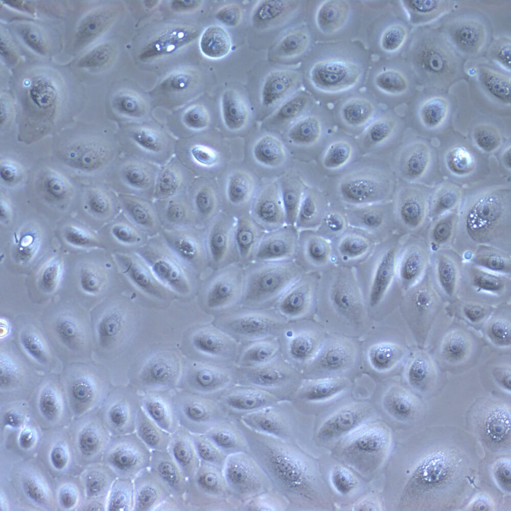
- Corneal epithelial, stromal, and endothelial cells
- Trabecular meshwork cells
- Iris pigment epithelial cells
- Retinal pigment epithelial cells
- Laminal cribrosal cells
- Choroidal fibroblasts
- Müller glia
If you don’t see the cell type you need, or have a specific project in mind, then contact us.
Frozen dissected globes
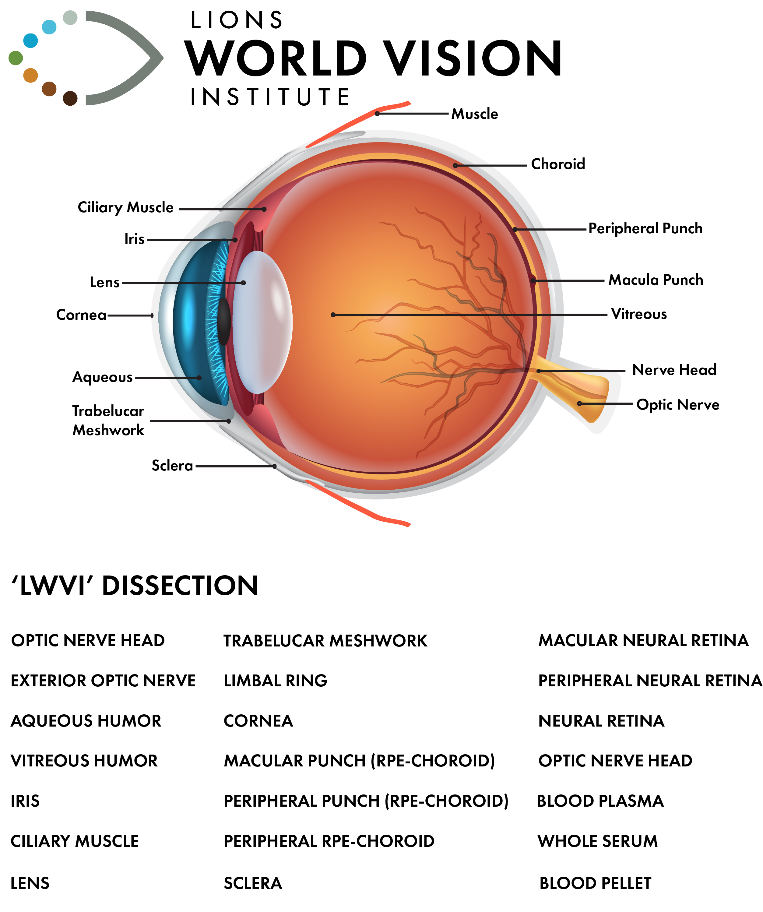
click to see moreThe “LWVI” dissection is our hallmark protocol, encompassing over 20 individual structures, humors, and blood samples to provide researchers with a comprehensive snapshot of ocular health and disease. This protocol, combined with grading for AMD and other pathologies, OCT imaging, and redacted medical histories, offers a robust framework for studying ocular conditions. These flash-frozen samples are an invaluable resource, ready for biomarker studies across a spectrum of human ocular diseases.
The LWVI utilizes the MGS grading system for grading AMD tissue, allowing us to provide unparalleled confidence in disease staging. Coupled with our rapid recovery times and the extensive repository, the LWVI is the world leader in providing high-quality ocular tissue to the world. For more information, please refer to our detailed grading system for AMD here.
Histological sections and fixed globes
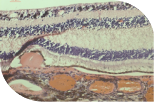
Paraffin Histology: AMD donor (200x Magnification)
with drusen
The LWVI also maintains a selection of clinically significant whole fixed globes and sections. With our complete histology suite and imaging facilities we can provide you with validation material for your project. We also offer staining and visualization optimization for brightfield and fluorescent imaging as well as expertise in ocular histology and handling. We can provide the full histological service from fixation to staining, imaging, and tissue microarray construction for advanced histology projects and next-generation applications such as spatial transcriptomics through our collaboration with the nearby Moffitt Cancer Institute. To request globes or sections, please contact us to discuss your project.